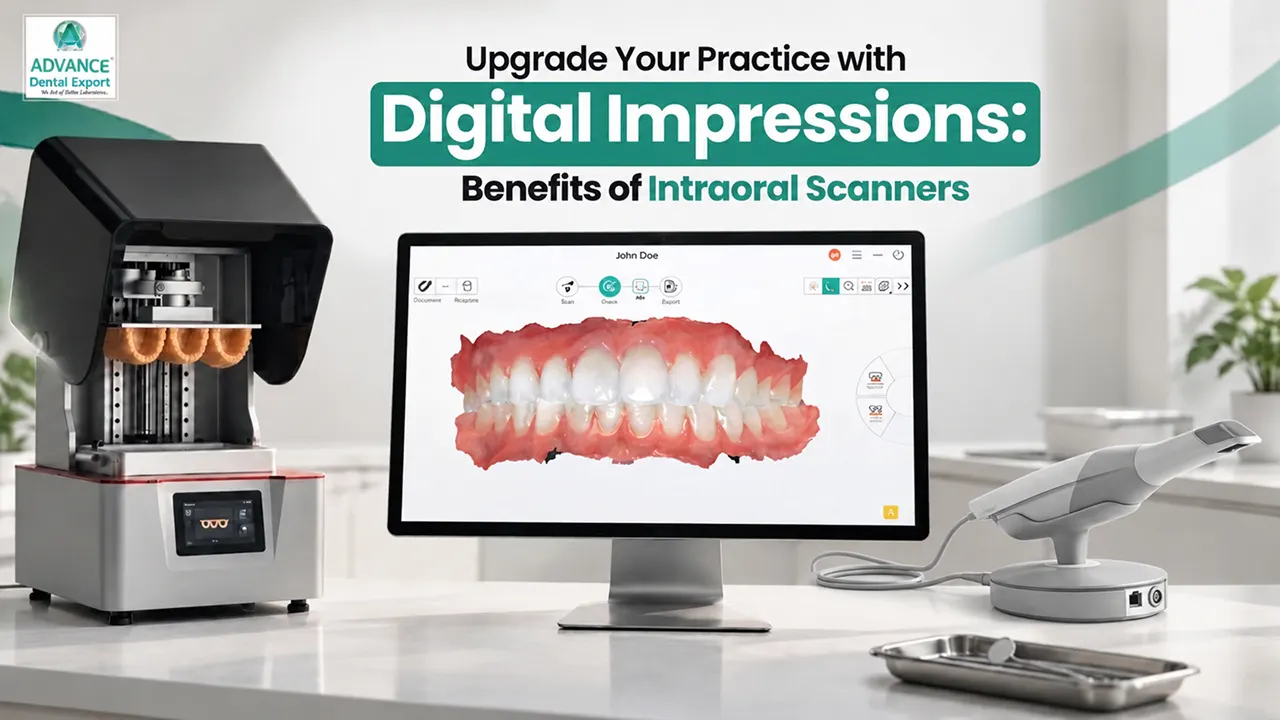
Digital impressions are changing how dentists capture dental anatomy, plan restorations, and communicate with dental laboratories. Instead of using conventional impression materials, dentists can use an intraoral scanner to create a detailed 3D digital model of the patient's teeth and surrounding oral structures.
These digital teeth impressions support CAD/CAM workflows for crowns, bridges, veneers, implant restorations, and orthodontic appliances.
For dental practices, digital scanning can improve patient comfort, reduce impression-related handling, and simplify laboratory communication. However, successful results depend on accurate scanning, proper tissue management, and case selection.
This guide explains how digital impressions work, their clinical applications, benefits, limitations, and practical integration into your dental practice.
What are Digital Impressions in Dentistry?
Digital impressions are three-dimensional (3D) records of a patient's teeth and accessible oral tissues, captured using an intraoral scanner or another compatible dental scanning system.
Digital impressions capture oral surfaces through optical scanning technology. The intraoral scanner converts the captured information into a virtual 3D model that dentists can examine, modify where appropriate, and share electronically with dental laboratories.
These models support computer-aided design and manufacturing (CAD/CAM), allowing dental technicians to design restorations digitally before milling or 3D printing suitable components.
Digital impressions are particularly useful for fixed prosthodontics, implant restorations, orthodontics, and aesthetic dental treatment planning.
What is an Intraoral Scanner?
An intraoral scanner (IOS) is a handheld digital dental scanning device that captures the visible surfaces of teeth, gingiva, and other accessible intraoral structures. It uses optical imaging technologies, such as structured light or confocal imaging, to collect surface information. Software processes overlapping images into a detailed 3D dental model.
The scanner displays the digital impression on a screen, allowing dentists to inspect preparations, identify missing areas, and rescan specific regions before submission. Common intraoral scanning systems include 3Shape TRIOS, iTero, and SHINING 3D Aoralscan.
Unlike a dental CBCT scanner, an intraoral scanner captures surface anatomy without ionising radiation. CBCT uses X-rays to image internal structures, including bone and roots, for appropriate diagnostic purposes.
How Do Digital Impressions Work Between Dentists and Dental Laboratories?
Digital impressions connect the dental clinic and laboratory through an electronic workflow, from capturing the patient's oral anatomy to designing and manufacturing the final restoration.
Step 1 : The process begins when the dentist captures a digital impression using an intraoral scanner. The scanning software creates a 3D model containing the preparation, adjacent teeth, opposing arch, and interocclusal relationship.
Step 2 : The dentist then sends the scan files and digital prescription to the dental laboratory through a compatible platform or secure file transfer.
Step 3 : Once the laboratory receives the case, technicians review the scan quality, preparation margins, interproximal contacts, and occlusal records.
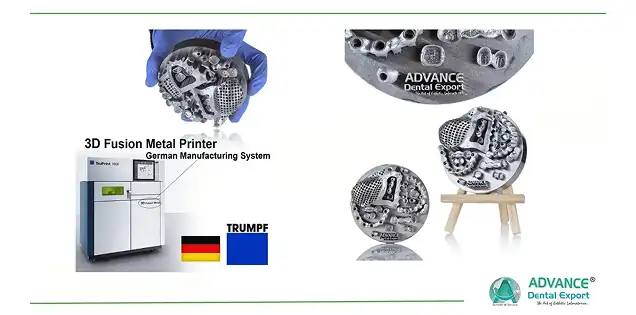
Step 4 : The approved scan data is imported into CAD software to design the restoration. Depending on the indication, the laboratory can manufacture restorations through CAD/CAM milling or use 3D printing for suitable models, patterns, guides, or other validated applications.
At Advance Dental Export (ADE) , we recognise that accurate digital impressions and complete prescriptions are essential for efficient communication between dentists and dental technicians.
The key consideration: A digital scan alone does not guarantee an accurate restoration. Clinical preparation, scan quality, material selection, and laboratory verification all influence the result.
What are the Clinical Applications of Digital Impressions?
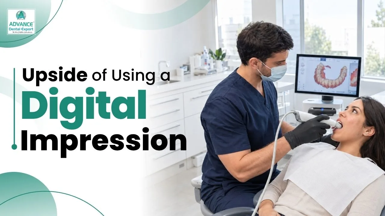
Digital impressions support restorative dentistry, prosthodontics, implantology, orthodontics, and aesthetic treatment planning.
Their clinical applications include:
- Crowns and bridges : Capture prepared teeth, finish lines, neighbouring contacts, and opposing occlusion for fixed restorations.
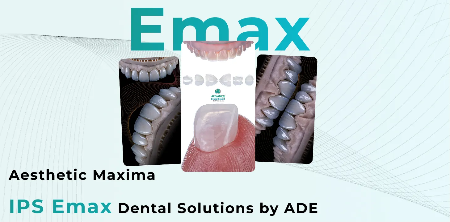
- Dental veneers, inlays, and onlays : Record tooth morphology and preparation details for CAD/CAM restorations.
- Implant-supported restorations : Capture implant positions using compatible scan bodies for digital prosthetic design.
- Orthodontics : Create digital study models, support clear aligner planning, and monitor tooth movement.
- Smile design : Combine 3D dental scanning with photographs and other records to plan aesthetic restorations.
- Occlusal appliances : Capture dental arches for digital splint and night guard fabrication.
Digital scanning also supports selected removable prosthodontic workflows.
How to Take Digital Impressions Using an Intraoral Scanner: Step-by-Step
Taking accurate digital impressions requires a systematic scanning technique, proper moisture control, and careful evaluation of captured surfaces.
Follow these steps to improve the quality of your intraoral scans.
Step 1: Prepare the Scanner and Patient
Select the appropriate scanning protocol and confirm that the scanner is ready. Reprocess reusable scanner tips according to manufacturer instructions.
Position the patient comfortably and maintain adequate access to the scanning area.
Step 2: Prepare the Scanning Area
Remove saliva, blood, and debris. Use isolation and gingival displacement where necessary to expose preparation margins.
Subgingival margins require particular attention because optical scanners cannot accurately record surfaces hidden beneath tissue.
Step 3: Scan the Prepared Arch
Follow the manufacturer's recommended scanning path. Capture the occlusal, buccal, and lingual or palatal surfaces.
Maintain the recommended scanning distance and movement speed. Ensure the preparation and adjacent contacts are completely recorded.
Step 4: Capture the Opposing Arch and Bite
Scan the opposing dentition and record the interocclusal relationship using the scanner's bite registration workflow.
Confirm that the virtual bite reflects the patient's intended intercuspal position. Investigate discrepancies before accepting the registration.
Step 5: Review the Digital Impression
Inspect the 3D model for missing surfaces, distorted anatomy, stitching errors, and unclear preparation margins.
Correct local defects through selective rescanning where appropriate. Do not rely on software to reconstruct clinically important missing anatomy.
Step 6: Complete and Submit the Case
Finalise the scan, enter the restoration prescription, attach relevant photographs, and submit the files through the laboratory's accepted platform.
Confirm that the prepared arch, antagonist, and bite registration are included before sending.
Research supports controlling moisture, following scanner-specific scanning strategies, and verifying captured data to improve impression accuracy.
Digital Impressions vs Traditional Dental Impressions: Which Is Better?
Digital impressions are often preferable for routine single crowns and short-span fixed restorations, while conventional impressions remain useful in complex clinical situations.
A comparison of both techniques helps dentists choose the appropriate workflow.
| Factor | Digital impressions | Traditional impressions |
| Technique | Optical 3D scanning | Impression trays and materials |
| Patient comfort | Generally better tolerated | May cause discomfort or gagging |
| Accuracy | Reliable for many short-span cases | Reliable with correct technique |
| Error correction | Selected areas can be rescanned | May require repeating the impression |
| Laboratory transfer | Electronic file submission | Physical impression or cast shipment |
| Initial investment | Scanner, software, and training | Lower equipment investment |
| Limitations | Hidden margins, soft tissues, extensive spans | Material distortion, tearing, and cast-related errors |
A 2026 overview of systematic reviews found no significant difference in marginal or internal fit for many fixed restorations.
Clinical takeaway: Choose digital or conventional impressions according to the indication, available equipment, operator proficiency, and restoration requirements.
What are the Benefits of Digital Impressions?
Digital impressions offer practical advantages for dentists, patients, and dental laboratories, especially when incorporated into an organised digital workflow.
Improved Patient Comfort
Digital scanning eliminates bulky impression trays and unpleasant impression materials. This can reduce gagging, nausea, and discomfort. Reviews have reported greater patient preference and comfort with intraoral scanning compared with conventional impressions.
More Efficient Clinical Workflows
Dentists can review scans immediately, correct selected areas, and send digital records without waiting for physical impression transportation.
Digital scanning may reduce procedure time, although efficiency depends on scanner performance, operator experience, and clinical complexity.
Better Dental Laboratory Communication
Digital models allow dentists and technicians to examine the same recorded anatomy.
This simplifies discussions about preparation margins, restoration design, occlusion, and other technical requirements before manufacturing.
Convenient Storage and Case Documentation
Digital impressions can be stored electronically and retrieved for treatment planning, follow-up comparisons, or future restorations.
However, digital accuracy still depends on proper clinical technique and complete records.
How to Integrate an Intraoral Scanner Into Your Dental Practice
Integrating an intraoral scanner requires selecting suitable equipment, training the dental team, establishing scanning protocols, and coordinating with your dental laboratory.
1. Choose the Right Intraoral Scanner
Select a scanner based on your clinical workload, restoration types, software requirements, and laboratory compatibility.
Common options include:
- 3Shape TRIOS : Offers digital restorative workflows and laboratory connectivity through 3Shape Unite.
- iTero intraoral scanners : Support compatible restorative and orthodontic digital workflows.
- Shining 3D Aoralscan : Supports digital impressions and exports common 3D file formats, including STL, OBJ, and PLY.
Compare scanning accuracy for your intended indications, ease of use, ergonomic design, supported file formats, local service availability, and recurring costs.
2. Evaluate the Intraoral Scanner Price in India
Intraoral scanner prices vary by brand, model, software package, and supplier.
Looking for a trusted lab partner? Get high-precision restorations from Advance Dental Export
Indian retailer listings include entry-level scanners at approximately ₹1.9–2 lakh and selected premium systems around ₹9.5 lakh. Other models require individual quotations, and these listings do not represent a fixed market-wide price range.
Consider the complete ownership cost, including software subscriptions, computer requirements, replacement tips, technical support, and maintenance.
3. Train Your Clinical Team
Begin with routine crown and short-span restorative cases before introducing complex digital workflows.
Train your team in scanner handling, moisture control, scanning sequences, bite registration, and quality assessment.
Develop a standard protocol for identifying incomplete scans and confirming patient records.
4. Coordinate With Your Dental Laboratory
Before submitting cases, confirm your laboratory's accepted file formats, prescription requirements, and digital communication platform.
STL files preserve 3D surface geometry, while formats such as PLY can also carry colour information. Check which data your laboratory needs for the intended restoration.
Establish a consistent process for reviewing scan problems, clarifying prescriptions, and securely transferring patient data.
A well-coordinated clinic–laboratory workflow is more valuable than purchasing a scanner without a defined implementation plan.
Conclusion
Digital impressions and intraoral scanners have become valuable tools for modern restorative and digital dentistry . They allow dentists to capture 3D dental records, improve patient comfort, and communicate efficiently with dental laboratories.
However, successful digital impressions depend on more than scanner technology. Accurate tooth preparations, tissue management, appropriate scanning techniques, and complete laboratory instructions remain essential.
For dentists in India, introducing digital scanning gradually and selecting equipment suited to clinical requirements can support a more consistent workflow.
Planning to integrate digital impressions into your dental practice? Connect with Advance Dental Export (ADE) to discuss your restorative case requirements and dental laboratory workflow.
FAQs Related to Digital Dental Impressions
What is a digital impression in dentistry?
A digital impression is a 3D record of a patient’s teeth and surrounding oral structures captured using an intraoral scanner. It provides digital data for designing crowns, bridges, veneers, and other dental restorations.
What is an intraoral scanner?
An intraoral scanner is a handheld digital device that uses optical imaging technology to capture the surfaces of teeth and surrounding oral tissues. It creates detailed 3D digital impressions without conventional impression materials or ionising radiation.
How much does an intraoral scanner cost?
Some intraoral scanners cost around ₹1.9–2 lakh in India, while premium systems cost around ₹9.5 lakh or more. Prices vary by brand, model, software subscriptions, accessories, and supplier.
What is the best dental scanner in 2026?
Popular intraoral scanning systems in 2026 include 3Shape TRIOS, iTero, and SHINING 3D Aoralscan. The best scanner depends on clinical requirements, scanning accuracy, software compatibility, laboratory integration, budget, and after-sales support.
How long does a digital impression take?
A digital dental impression typically takes a few minutes, depending on the scanning area, case complexity, and scanner used. Dentists can immediately review the 3D model, identify missing details, and rescan specific areas when necessary.
What is a 3D scan in dentistry?
A 3D dental scan creates a three-dimensional digital representation of dental structures. Intraoral scanners capture visible tooth and gum surfaces for restorative planning, while CBCT scans use X-rays to visualise internal structures such as bone and tooth roots.
Does Advance Dental Export accept digital impressions?
Yes. Advance Dental Export (ADE) accepts digital impressions and supports CAD/CAM workflows for crowns, bridges, veneers, and implant restorations. You can submit compatible STL files through ADE's online submission portal.
Does ADE provide intraoral scanning services?
Yes, Advance Dental Export (ADE) offers intraoral scanning services across many cities in India. Availability of chairside or on-site scanning for individual dental clinics should be confirmed directly with ADE. Contact us.